What Is Peroneal Tendinosis?
The peroneal tendons are on the outside of the ankle just behind the bone called the fibula. Peroneal tendinosis is the name for the enlargement, thickening, and swelling of these tendons. This usually occurs with overuse, such as a repetitive activity that irritates
the tendon over long periods of time.
Symptoms
People with peroneal tendinosis typically have tried a new exercise or markedly increased their activities. Characteristic activities include marathon running or others that require repetitive use of the ankle. Patients usually have pain around
the back and outside of the ankle. There usually is no history of a specific injury.
Causes
Improper training or rapid increases in training and poorly fitting shoes can lead to peroneal tendinosis. Also, patients who have high arches may be more susceptible because their heel is turned inwards slightly, which requires the peroneal tendons to work
harder to turn the ankle to the outside. The harder the tendons work, the more likely patients are to develop tendinosis.
Anatomy
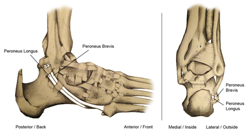
Tendons connect muscle to bone and allow them to exert their force across the joints that separate bones. Ligaments, on the other hand, connect bone to bone. There are two peroneal tendons that run along the back of the fibula. The first is called the
peroneus brevis. The term "brevis" implies short. It is called this because it has a shorter muscle and starts lower in the leg. It then runs down around the back of the bone called the fibula on the outside of the leg and connects
to the fifth metatarsal on the side of the foot.
The peroneus longus takes its name because it has a longer course. It starts higher on the leg and runs all the way underneath the foot to connect to the first metatarsal on the other side. Both tendons share the major job of turning
the ankle to the outside. The tendons are held in a groove behind the back of the fibula and are covered by a ligament-type tissue called a retinaculum.

Diagnosis
Your foot and ankle orthopaedic surgeon will take your history and perform an exam to make the diagnosis. Most patients with peroneal tendinosis will report overuse of activity, rapid increase in recent activity, or other training errors, along with pain
in the back and outside of the ankle. During the exam, you may feel pain when the surgeon touches the peroneal tendons.
It is important to distinguish this from pain over the fibula, which might indicate a different problem. Pain on the fibula occurs directly over the bone. Pain in the peroneals occurs slightly further behind. You may also have pain when you turn the foot inside towards the middle of your body (inversion) and weakness when you try to bring the foot to the outside (eversion). Your surgeon also will look for varus alignment of the heel,
which means that the heel is turned inwards.
X-rays for peroneal tendinosis usually are normal. Ultrasound is a very effective way to assess the tendons and can show an abnormal appearance or tear.
An MRI also may show a tear.
Treatments
Non-Surgical Treatment
The vast majority of peroneal tendinosis cases will heal without surgery. This is because it is an overuse injury and can heal with rest. If there is significant pain, wearing a CAM walker boot for several weeks is a good idea. If there is no tenderness
with walking, an ankle brace might be the next best step. You should limit how much you are walking or standing until the pain improves. This usually takes several weeks. After this period you can resume training, but very slowly
and based on pain. For patients with varus alignment, an orthotic that tilts the ankle to the opposite side may help relieve pressure on the tendons. It is important to talk to your doctor about changing your training. This includes using new shoes for running or cross-training instead. Physical therapy also is
very important to strengthen the tendons.
There is increasing use of platelet-rich plasma (PRP) and stem cell injections to assist with healing; however, the research on this is limited. Steroid injections are not routinely performed due to the risk of causing complete rupture.
Surgical Treatment
Your foot and ankle orthopaedic surgeon may recommend surgical treatment if the pain continues. Non-surgical treatment could last up to a year before considering surgery. If there is a tear, meaning a split that runs along the length of the tendons, your surgeon could consider repairing the tendon. Sometimes, making the groove in the back of the bone of the fibula deeper allows the tendons more space and can help as well. Finally, if the tendon is very diseased, your surgeon may need to resect the tendon and connect
both the longus and brevis together. Only the specific tendon involved should be addressed. Occasionally, both may be involved and more complex reconstruction necessary.
Recovery
Patients usually recover fully but this can take considerable time. You must be patient and allow the tendon to heal before going back to activity. If you need surgery, your recovery time may be substantial. You may be instructed not to bear weight for
about six weeks. Your foot and ankle orthopaedic surgeon likely will order physical therapy once you're ready.
Risks and Complications
In the case of surgery, as for all surgeries, there is a risk of developing an infection or wound complication. Nerve damage can occur, in particular the sural nerve, which runs along the outside of the ankle near the tendons and provides sensation to the outside of the foot.
What would happen if my peroneal tendinosis is not treated?
If the tendinosis is not addressed, your tendon can tear. Also, weakness of the tendons can lead to an ankle sprain or chronic ankle instability, which can cause damage to the cartilage inside the ankle joint.
What is the difference between tendinitis and tendinosis?
With tendinitis, there is acute inflammation of the tendons. This usually happens directly after an injury for a short period of time. Tendinosis is a chronic process
of pain in the tendons that results in an enlargement and thickening of the tendon.
Original article by Scott Holthusen, MD
Reviewers/Contributors: David Garras, MD; Raymond Walls, MD
The American Orthopaedic Foot & Ankle Society (AOFAS) offers information on this site as an educational service. The content of FootCareMD, including text, images, and graphics, is for informational purposes only. The content is not intended to substitute
for professional medical advice, diagnoses or treatments. If you need medical advice, use the "Find a Surgeon" search to locate a foot and ankle orthopaedic surgeon in your area.

